
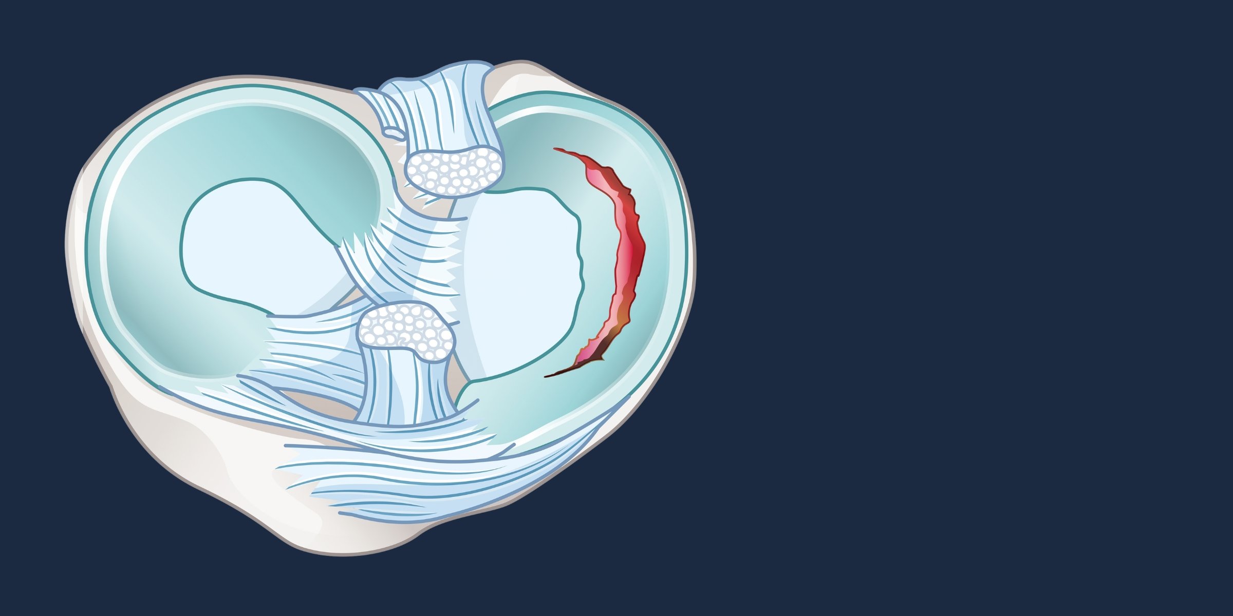
A meniscal tear is the most common serious injury to the knee. Most people who are told they have a "torn cartilage" have a meniscal tear. Not all meniscal tears are the same, however - and the right treatment depends on the type of tear, the patient's age, and the overall state of the knee.
What Is the Meniscus?
The knee has two menisci; the medial meniscus on the inner side of the knee, and the lateral meniscus on the outer side. Each is a C-shaped wedge of fibrocartilage that sits between the end of the thighbone (femur) and the top of the shinbone (tibia).
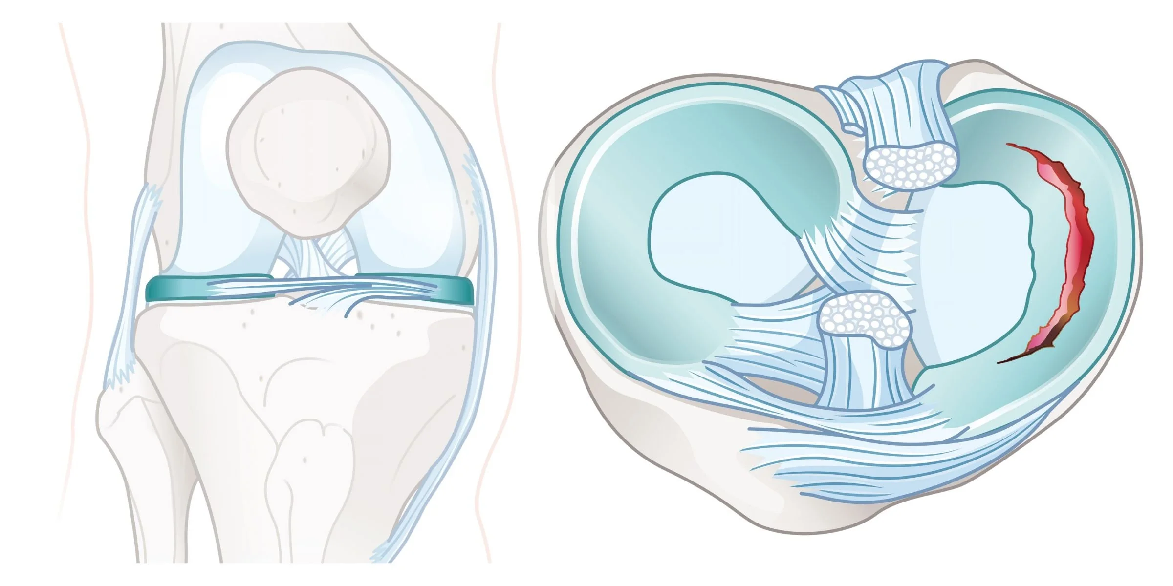
Left: the knee from the front, with the menisci (teal) sitting between the thighbone and shinbone. Right: looking down on the top of the shinbone. The lateral meniscus is on the left and the medial meniscus on the right, with a tear shown in red.
The menisci serve several important functions. They distribute load evenly across the knee joint, absorb shock during walking and activity, and contribute to the stability of the joint. They also help distribute fluid within the joint, assisting with lubrication of the articular cartilage.
The outer third of each meniscus has a blood supply - this is called the vascular zone. The inner two-thirds have no blood supply. This distinction matters for treatment: tears within the vascular zone have the potential to heal; tears in the inner avascular zone do not.
Types of Meniscal Tears
Meniscal tears are broadly divided into two categories: traumatic tears and degenerative tears. These differ in how they occur, who they affect, and how they are best managed.
Traumatic Tears
Traumatic tears occur as a result of an acute injury - typically a twisting or pivoting movement on a loaded knee. They are more common in younger, active people. A common presentation is an athlete who twists their knee during sport, feels immediate pain and swelling, and has difficulty returning to activity.
Traumatic tears often occur alongside other injuries. A tear of the medial meniscus frequently accompanies an ACL rupture, because both structures are stressed by the same mechanism of injury.
Tear patterns vary. A bucket handle tear - where a large segment of the meniscus tears in a semicircular shape and folds into the joint - can cause the knee to lock, preventing full straightening. Radial tears, flap tears, and root tears are other patterns that may occur with acute injury. The specific pattern influences whether the tear can be repaired or needs to be partially removed.
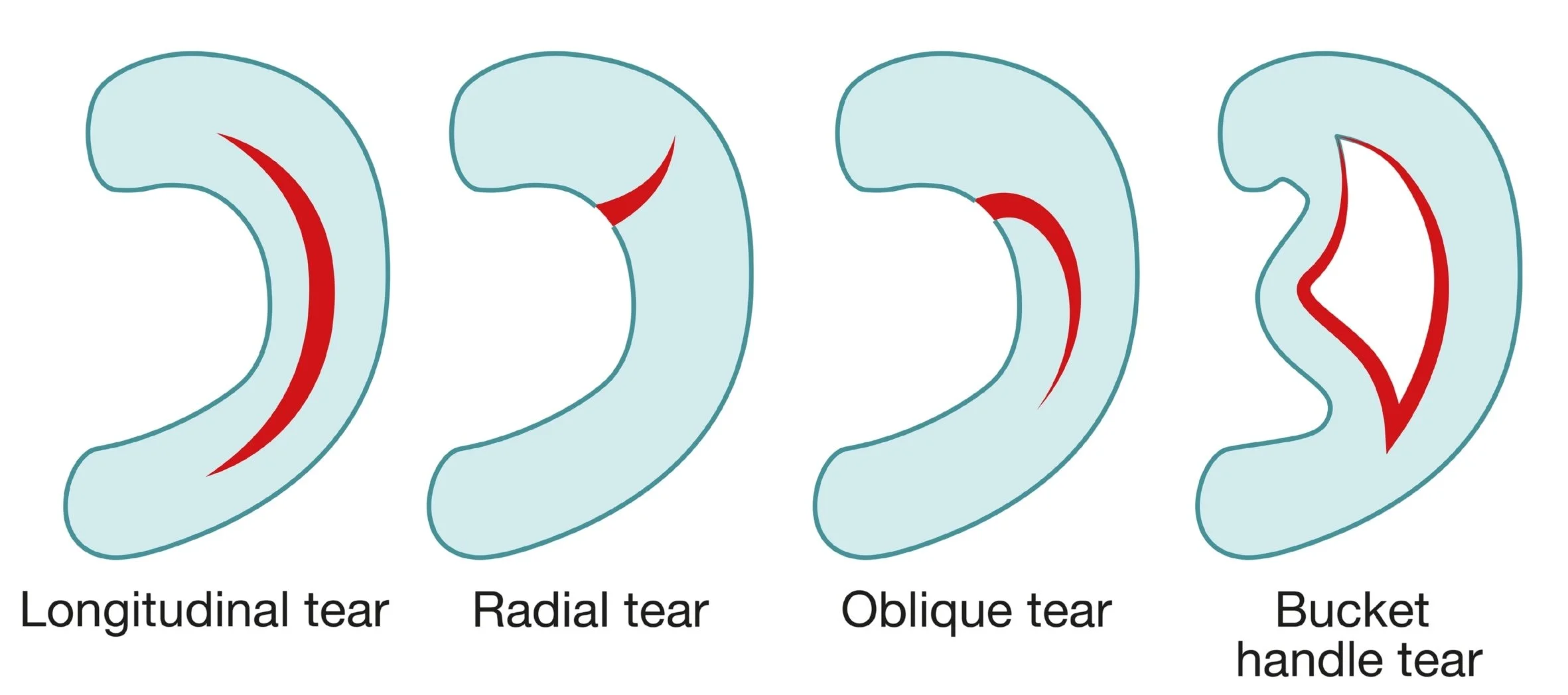
Common meniscal tear patterns, seen from above, with the tear shown in red. Oblique tears are also known as flap or parrot-beak tears.
Degenerative Tears
Degenerative tears occur as part of the natural ageing process of the knee. They are common in people over 40 and often occur without a clear injury, sometimes from something as minor as rising from a chair or stepping awkwardly. In many cases, there is no identifiable triggering event at all.
Degenerative meniscal tears are closely associated with early osteoarthritis. The meniscal tissue becomes less resilient over time and is more susceptible to tearing under normal loads. In people over 50, meniscal changes are common findings on MRI - many of which cause no symptoms at all.
This is an important distinction when it comes to treatment. The presence of a meniscal tear on an MRI scan does not automatically mean that the tear is the source of pain. Clinical examination and correlation with the patient's symptoms are essential before any treatment decision is made.
Symptoms
The symptoms of a meniscal tear depend on the type, size, and location of the tear. Common symptoms include:
Pain along the inner or outer joint line of the knee (depending on which meniscus is involved)
Swelling; which may be immediate after an acute tear, or may develop gradually over 24–48 hours
Clicking, catching, or a sensation of something moving within the joint
Difficulty fully bending or straightening the knee
Pain with squatting, pivoting, or prolonged walking
A sense of the knee giving way or feeling unstable
A knee that is locked (unable to be straightened fully) is a specific symptom that suggests a displaced bucket handle tear. This is a situation that typically requires prompt surgical assessment.
Diagnosis
Diagnosis of a meniscal tear begins with a clinical examination. Tenderness along the joint line, combined with specific tests that load the meniscus under rotation, can identify a likely tear in most cases. Clinical examination also helps distinguish between a meniscal tear and other causes of knee pain, including ligament injury, articular cartilage damage, and early arthritis.
X-rays do not show the meniscus, but they are useful for assessing the overall alignment and joint space of the knee and for identifying signs of osteoarthritis.
MRI is the primary imaging investigation for a suspected meniscal tear. It provides detailed information about the size, pattern, and location of the tear, and also assesses the articular cartilage, ligaments, and bone. MRI has high sensitivity and specificity for meniscal pathology.
It is worth noting that MRI findings must always be interpreted in the context of clinical examination. In people over 40, changes within the meniscus on MRI are common and are not always the cause of symptoms. A decision about treatment is based on the combination of clinical assessment and imaging - not on the MRI report alone.
Treatment Options
Treatment depends on the type and pattern of the tear, the patient's age and activity level, and the overall condition of the knee joint.
Non-Surgical Management
For degenerative meniscal tears, non-surgical management is the recommended first-line treatment. A structured physiotherapy programme (focusing on strengthening the muscles around the knee, improving neuromuscular control, and managing load) produces good outcomes in the majority of patients.
Current evidence shows that, for degenerative tears in middle-aged patients without significant mechanical symptoms, outcomes following physiotherapy are equivalent to those following surgery. A thorough trial of non-surgical management (typically at least eight to twelve weeks) is appropriate before surgical options are considered.
Not all patients respond equally to non-surgical management. Those with true mechanical symptoms - reproducible catching, locking, or instability attributable to the tear - may have a faster or more complete recovery following surgery, and several studies support a surgical approach in this subgroup. The treatment decision is made on an individual basis after full assessment.
Anti-inflammatory medication and activity modification may help manage symptoms during rehabilitation. Intra-articular corticosteroid injection can provide short-term relief in some patients, particularly where there is associated synovitis.
Arthroscopic Partial Meniscectomy
Arthroscopic partial meniscectomy (APM) is the surgical removal of the torn portion of the meniscus, leaving as much healthy meniscal tissue as possible. It is performed arthroscopically through two or three small incisions, typically as a day procedure under general anaesthetic.
The procedure is appropriate when mechanical symptoms (locking, persistent catching, or instability) are present and attributable to the tear, and for patients in whom an adequate trial of non-surgical treatment has not produced satisfactory improvement.
Recovery after APM is generally straightforward. Most patients are walking normally within one to two weeks and return to light activities within two to three weeks. Return to sport is typically possible by six to eight weeks, depending on the nature of the activity and the extent of the resection.
Meniscal Repair
Where a tear occurs in the outer vascular zone of the meniscus, and the tear pattern is suitable, it may be possible to repair the meniscus rather than remove the torn tissue. Repair is preferred over resection wherever it is technically feasible, because it preserves meniscal tissue and its long-term protective effect on the knee joint.
Meniscal repair is performed arthroscopically, using sutures or implants to bring the torn edges together and allow biological healing. It is more commonly performed in younger patients with acute traumatic tears in the vascular zone. Bucket handle tears are often suitable for repair.
Recovery after meniscal repair is longer than after partial meniscectomy, because the repair must be protected while healing occurs. Weight-bearing may be restricted for four to six weeks, and return to pivoting or cutting activities typically takes three to four months.
When Is Surgery Appropriate?
Surgery is most clearly indicated in the following situations:
A locked knee - where a displaced bucket handle tear is preventing full extension
A traumatic tear in a younger, active patient with persistent mechanical symptoms that have not responded to rehabilitation
A tear associated with ACL reconstruction, where the meniscal injury is addressed at the time of the ligament procedure
A degenerative tear in which an adequate trial of non-surgical management has not produced satisfactory improvement
A tear in the vascular zone that is suitable for repair, particularly in a younger patient
The decision to proceed with surgery is made after full assessment; taking into account the type of tear, the patient's symptoms and functional demands, the response to non-surgical treatment, and the findings on MRI. There is no single correct answer for every patient, and the approach is individualised.
Recovery
Recovery depends primarily on what procedure was performed. After arthroscopic partial meniscectomy, the majority of patients are comfortable within the first week and return to normal daily activities within two to three weeks. Swelling settles progressively over four to six weeks. Return to sport is typically at six to eight weeks, with a graduated return to full activity.
After meniscal repair, recovery is more gradual. The repaired meniscus must be protected to allow healing. Most patients use crutches for the first four to six weeks and avoid deep flexion and twisting during this early healing period. Return to sport is expected at three to four months, with full recovery at six months for some repair patterns.
Physiotherapy is important after either procedure - to restore range of motion, rebuild muscle strength, and support a safe return to activity.
Key Points
The tear type matters
Traumatic tears in younger patients and degenerative tears in middle-aged patients behave differently and are treated differently. The right approach depends on which type is present.
Non-surgical management first for degenerative tears
Current evidence supports physiotherapy as the recommended first-line treatment for degenerative meniscal tears. Surgery has a clear role, however, when an adequate trial of non-surgical treatment has not produced satisfactory improvement.
MRI findings require clinical correlation
Meniscal changes on MRI are common in people over 40 and are not always the source of symptoms. A positive MRI finding must be interpreted alongside clinical examination before any treatment decision is made.
Repair is preferred over resection where possible
When a tear is in the vascular zone and the pattern is suitable, repair is preferred over partial meniscectomy because it preserves the meniscal tissue and its protective function within the knee.
A locked knee warrants prompt assessment
Inability to fully straighten the knee after a meniscal injury suggests a displaced bucket handle tear. This is a specific situation that requires timely orthopaedic review.
The information provided on this page is for general educational purposes and does not constitute medical advice. If you have knee pain or a suspected meniscal injury, please seek assessment from a qualified medical practitioner. Treatment recommendations are made on an individual basis following full clinical assessment.