
A shoulder dislocation (where the ball of the upper arm comes out of the socket) is one of the most common joint injuries seen in active people, particularly in contact sport and in young adults. For many people, a single dislocation that is reduced and rehabilitated is the end of the story. For others, the shoulder continues to dislocate or feels persistently unstable, affecting confidence, activity, and quality of life.
Understanding why some shoulders become unstable after a dislocation - and what can be done about it - is the focus of this article.
How the Shoulder Joint Works
The shoulder is the most mobile joint in the body. It is a ball-and-socket joint: the ball is the head of the humerus (the upper arm bone), and the socket is the glenoid, which is part of the shoulder blade. Unlike the hip - which has a deep, well-contained socket - the glenoid is relatively shallow and flat. This shallowness is what gives the shoulder its exceptional range of movement, but it also means the joint relies heavily on the surrounding soft tissues for stability.
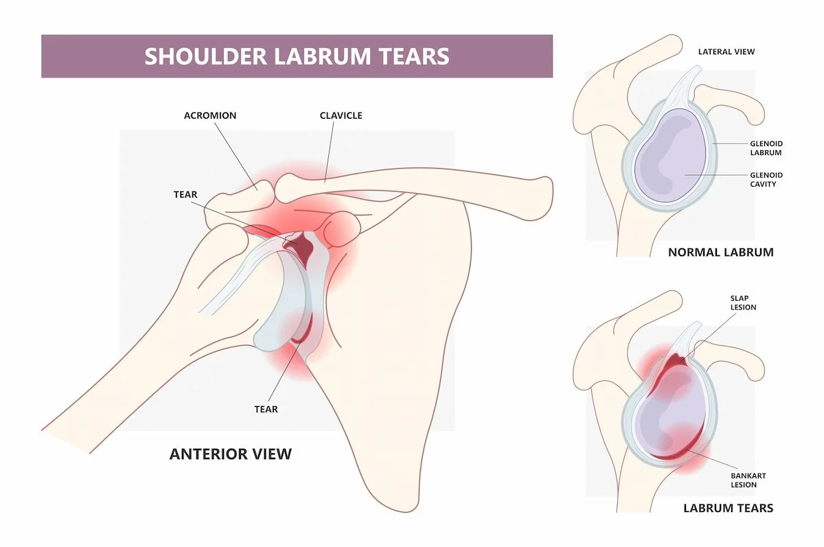
The primary stabilising structure is the labrum - a ring of fibrocartilage that surrounds the rim of the socket, deepening it and providing an anchor point for the ligaments that hold the joint together. The rotator cuff muscles provide dynamic stability, and the glenohumeral ligaments provide passive restraint at the limits of movement.
What Happens When the Shoulder Dislocates
The vast majority of shoulder dislocations are anterior; the ball comes out of the front of the socket. This usually happens when the arm is forced into abduction and external rotation, such as during a tackle, a fall onto an outstretched arm, or a collision.
At the moment of dislocation, the labrum is torn away from the front of the socket. This is called a Bankart lesion, and it occurs in the majority of first-time traumatic dislocations. The force of the ball striking the rim of the socket as it comes out also often creates a dent in the back of the ball itself; a Hill-Sachs lesion. In some cases, a fragment of bone is avulsed from the rim of the socket, which is called a bony Bankart lesion.
These are structural injuries. The labrum does not reliably heal back to its original position after a dislocation, and the Hill-Sachs and bony Bankart lesions represent permanent changes to the bony architecture of the joint. This is why the shoulder can remain vulnerable to further dislocation even after the acute episode has settled.
Why Some Shoulders Keep Dislocating
After a first dislocation, the risk of recurrence varies considerably depending on age and activity level. In patients under 25, particularly those who play contact or overhead sport, recurrence rates after conservative management alone are reported as high as 70 to 90% in the published literature. In older patients with lower activity demands, the risk is substantially lower.
The reasons for this are partly structural and partly behavioural. The labrum has not healed, the ligaments have been stretched, and the bony architecture may have changed. A young person returning to rugby, football, or any sport involving overhead or contact activity is repeatedly loading a joint that has lost some of its normal stabilising mechanism.
Some patients develop ongoing instability without repeated full dislocations; a sense that the shoulder is about to come out, or that it slips partially out and back in (subluxation). This can be equally limiting and is part of the same spectrum of instability.
Symptoms
The symptoms of shoulder instability vary depending on whether the shoulder has dislocated once or repeatedly, and how much the underlying anatomy has been affected.
Common symptoms include:
A history of one or more shoulder dislocations requiring reduction
A feeling that the shoulder is about to come out with certain movements - particularly reaching overhead, behind the head, or into external rotation
Apprehension with arm positions that previously caused a dislocation
Shoulder pain with overhead activity or contact sport
A sense of weakness or lack of trust in the shoulder during activity
In some cases, a clunk or catch as the shoulder moves
Diagnosis
The diagnosis of shoulder instability is primarily clinical; based on the history of dislocation, the pattern of symptoms, and examination findings including the apprehension test and relocation test.
Imaging plays an important role in understanding the extent of the structural damage:
Plain x-ray (AP shoulder and axillary view) - requestable by your GP. The axillary view in particular provides information about the shape of the glenoid and may identify a bony Bankart fragment or significant Hill-Sachs lesion. An x-ray does not show the soft tissue labrum but is an important first step.
MRI arthrogram - the most accurate investigation for assessing the labrum and associated structures. Contrast is injected into the joint before the scan, which significantly improves the visibility of labral tears and ligamentous injuries. In New Zealand, an MRI arthrogram is arranged through the orthopaedic specialist after referral rather than in primary care.
CT scan - used when significant bone loss is suspected, to quantify the size of a bony Bankart lesion or Hill-Sachs defect. This information directly informs surgical planning.
In patients over 40, a first dislocation is more likely to be associated with a rotator cuff tear in addition to labral damage. MRI is particularly important in this age group to identify any concurrent rotator cuff injury.
Treatment
Treatment depends on the severity of the instability, the extent of the structural damage, the patient's age and activity level, and whether this is a first or recurrent episode.
Conservative Management
After a first dislocation, the standard initial approach is a period of immobilisation in a sling followed by a structured physiotherapy programme. Rehabilitation focuses on rebuilding the strength and neuromuscular control of the rotator cuff and periscapular muscles to compensate for the structural damage and reduce the risk of further dislocation.
For many patients, particularly those who are older or less active, physiotherapy is sufficient to manage symptoms and return to normal activity. Conservative management is the appropriate starting point in most cases.
When Surgery May Be Considered
Surgery is considered when:
The shoulder has dislocated on more than one occasion and conservative management has not provided adequate stability
A first dislocation has occurred in a young, active patient with a high risk of recurrence who wishes to return to contact or high-demand sport; in selected cases, early surgical stabilisation may be discussed
There is significant bone loss on imaging (a large Hill-Sachs lesion or bony Bankart) that is likely to cause recurrent instability
The decision to proceed to surgery is made on an individual basis, taking into account the imaging findings, the pattern of instability, the patient's goals, and the risks and benefits of the procedure.
Surgery for Shoulder Instability
There are two main surgical options for shoulder instability. The choice between them depends primarily on whether significant bone loss is present.
Bankart Repair
When the labrum has torn but the bony anatomy of the joint is intact, a Bankart repair is the standard procedure. This is performed arthroscopically (through small keyhole incisions) under general anaesthetic.
Using a camera and small instruments inside the joint, the torn labrum is reattached to the front of the glenoid rim using suture anchors (small implants that hold the repair in place while it heals). The ligaments are tensioned at the same time. The procedure typically takes around 45 to 60 minutes and is done as a day case.
Where a significant engaging Hill-Sachs lesion is present, a Remplissage procedure may be performed at the same time as the Bankart repair. Remplissage - from the French for filling - involves anchoring a portion of the posterior capsule and the infraspinatus tendon into the Hill-Sachs defect, effectively filling it and preventing it from engaging on the glenoid rim during movement. It is performed arthroscopically as part of the same operative session and does not significantly alter the recovery timeline.
A sling is worn for four to six weeks following surgery. Physiotherapy begins early, with gentle range of motion in the first few weeks and progressive strengthening from around six weeks. Return to contact sport is typically around six months after surgery.
Latarjet Procedure
When there is significant bone loss from the glenoid - a bony Bankart lesion or a shallow, deficient socket - a Bankart repair alone may not restore adequate stability. In these cases, the Latarjet procedure is used.
The Latarjet involves transferring a small piece of bone (the coracoid process) from the front of the shoulder blade to the front of the glenoid, where it is fixed with screws. This has two effects: it rebuilds the bony rim of the socket, and the conjoint tendon attached to the coracoid provides a dynamic sling effect that resists anterior translation of the humeral head.
It is an open procedure, more extensive than a Bankart repair, and is used in specific circumstances rather than as a routine approach. Recovery follows a similar timeline: sling for four to six weeks, structured rehabilitation, and return to contact sport at around six months.
Recovery After Surgery
The general recovery milestones following surgery for shoulder instability are:
Sling: four to six weeks
Driving: typically from four to six weeks once the sling is removed and comfortable
Return to desk work or light activity: two to four weeks in most cases
Return to overhead activity and gym work: from three to four months
Return to contact sport: from six months
The tissue healing process takes time regardless of the procedure performed. Physiotherapy throughout the rehabilitation period is important for achieving the best result. Full confidence in the shoulder - the feeling of trust that it will not come out - often returns gradually over the months following surgery as strength and proprioception improve.
When to Seek an Orthopaedic Opinion
It is reasonable to seek an orthopaedic opinion if:
The shoulder has dislocated more than once
There is persistent apprehension or instability despite physiotherapy
You are a young, active person who has had a first dislocation and wants to understand the options
Imaging has shown significant bone loss or a bony Bankart lesion
A consultation with your GP or physiotherapist is the appropriate first step. Your GP or physiotherapist will be able to request a shoulder x-ray and arrange an ACC claim if the dislocation occurred as a result of a traumatic event, which affects the funding pathway for any subsequent treatment.
Key Points
-
The ball-and-socket design of the shoulder prioritises movement over stability, making the labrum (a ring of cartilage around the socket) essential for keeping the joint in place.
-
A traumatic dislocation typically tears the labrum from the front of the socket (a Bankart lesion) and may also dent the back of the ball (a Hill-Sachs lesion). Without repair, recurrence rates are high - particularly in young, active patients.
-
Initial treatment is sling immobilisation followed by physiotherapy. Surgery may be considered after recurrent instability or in selected high-risk patients after a first dislocation.
-
Arthroscopic surgery reattaches the torn labrum to the glenoid rim using small anchors. Where a significant engaging Hill-Sachs lesion is present, a Remplissage procedure may be added to the procedure.
-
When there is significant glenoid bone loss, a Bankart repair alone is insufficient. The Latarjet procedure transfers a bone block to the front of the socket to restore stability - it is an open procedure with a longer recovery.
This article is for general educational purposes only and does not constitute medical advice. Please consult your doctor or seek a specialist referral for personal medical advice.