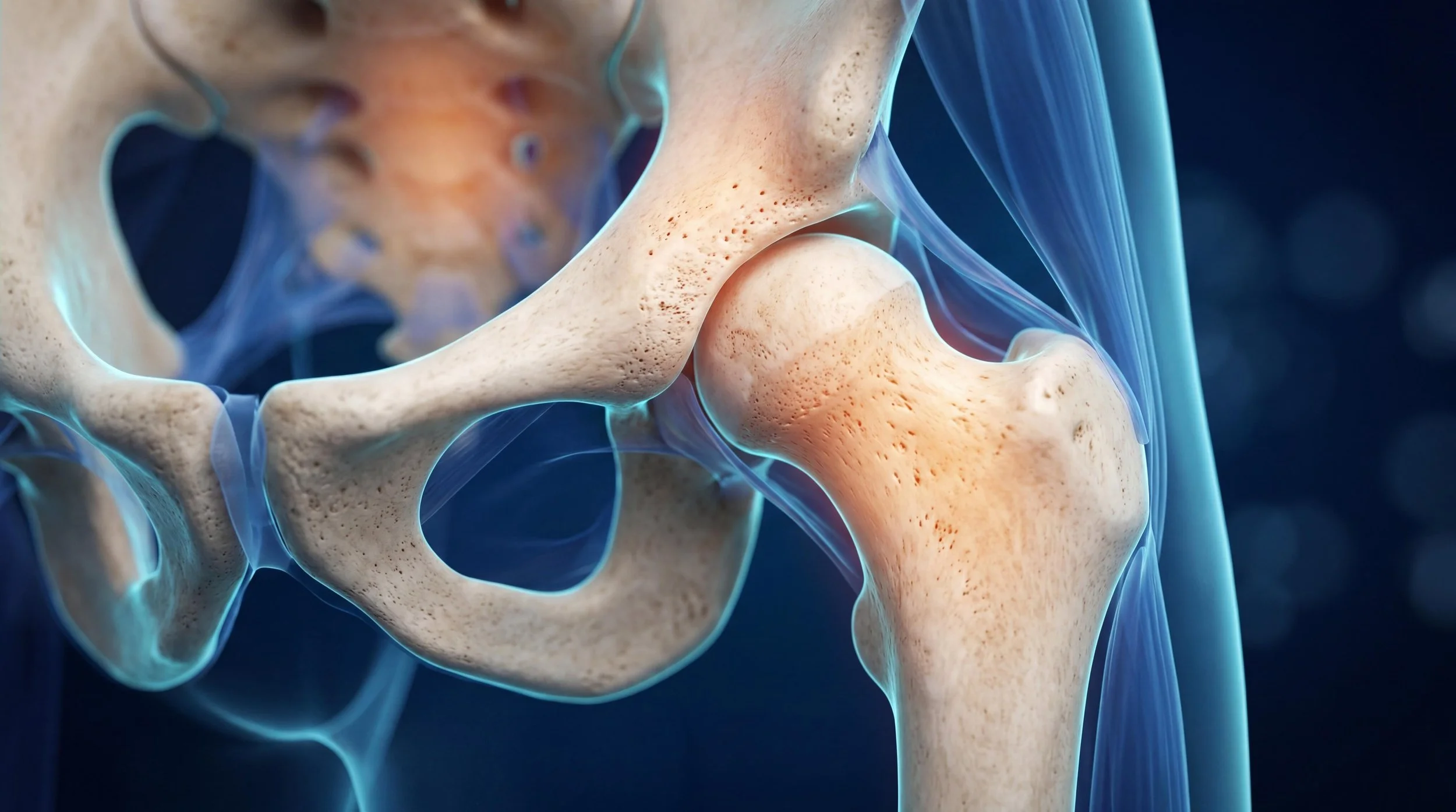
Hip

PAO Surgery — A Complete Guide for Patients with Hip Dysplasia

Periacetabular osteotomy — PAO — is the most effective surgical treatment available for young adults with hip dysplasia, and it is an operation that very few surgeons in New Zealand perform. For patients who have spent years searching for the source of their hip pain, understanding what PAO involves, who it is suitable for, and what recovery looks like is an important step in making an informed decision about their care.

For a video explanation of what hip dysplasia is and how it is diagnosed, see our Hip Dysplasia guide.

What Is PAO Surgery?

PAO is a joint-preserving hip surgery in which the acetabulum (the hip socket) is surgically cut free from the pelvis, repositioned to provide better coverage of the femoral head (the ball of the hip joint), and fixed in its new position with screws. By improving the coverage and orientation of the socket, PAO redistributes load more evenly across the cartilage surface, reduces pain, protects the labrum, and — in appropriately selected patients — significantly delays or prevents the progression to hip arthritis and the need for total hip replacement.

The procedure was developed in the 1980s and has been refined over decades of clinical experience. Long-term follow-up studies consistently show that PAO produces excellent results in appropriately selected patients, with many patients maintaining their native hip joint into their fifties and sixties.

Who Is a Candidate for PAO?

PAO is most appropriate for:

  • Patients with symptomatic hip dysplasia — groin pain, labral tears, or hip instability attributable to inadequate acetabular coverage

  • Patients with preserved or near-preserved joint cartilage — PAO is a joint-preserving operation, not a salvage procedure; if significant arthritis has already developed, the benefit is substantially reduced

  • Patients typically aged between 15 and 45 years, though the upper age limit depends on cartilage and bone quality and is assessed individually

  • Patients who have not obtained adequate symptom relief from non-operative management

PAO is not appropriate for patients with severe arthritis, patients with other significant hip deformity that requires different surgical correction, or patients whose symptoms are not primarily attributable to dysplasia.

A careful preoperative assessment — including detailed imaging and clinical examination — is essential to confirm that PAO is the right operation for each individual patient.

What Does PAO Surgery Involve?

PAO is performed under general anaesthetic and takes approximately 2–3 hours. Through an incision over the front of the hip, the surgeon makes precise cuts around the acetabulum to free it from the surrounding pelvis. The acetabulum is then carefully repositioned — typically redirected laterally, anteriorly, and slightly medially — to improve the coverage of the femoral head. Once optimal position is confirmed under intraoperative imaging, the repositioned socket is secured with three or four stainless steel screws.

In some cases, hip arthroscopy is performed in the same sitting to assess and treat any labral tears or cartilage damage within the joint.

What Does Recovery from PAO Look Like?

Recovery from PAO is a significant undertaking — it is a major pelvic reconstruction, and both patients and their families should be prepared for a meaningful period of rehabilitation.

Hospital stay: 4–5 days in hospital following surgery.

Weeks 1–6: Protected weight-bearing with crutches. The screws allow early mobilisation, but the healing bone requires protected loading during this phase. Physiotherapy begins in hospital and continues after discharge.

6–12 weeks: Gradual increase in weight-bearing as directed by the surgeon and physiotherapist. Walking without crutches typically begins during this phase.

Months 3–6: Progressive strengthening and return to everyday activities. Most patients return to desk work or study within 4–8 weeks. Driving is typically possible by 6–8 weeks (for an automatic vehicle) once off crutches and comfortable.

Months 6–12: Return to lower-impact sport and recreational activity.

12+ months: Return to more demanding physical activities and sport, guided by objective assessments.

The screws are not routinely removed but can be taken out as a minor procedure if they cause symptoms.

Why Seek Care at a Specialist Centre?

PAO is a technically demanding operation that requires specific training and ongoing case volume to maintain proficiency. Outcomes are strongly correlated with surgeon experience. Dr. Matthew Boyle trained in PAO surgery during his fellowship at Harvard University — one of the world’s leading hip preservation centres — and has performed PAO for patients from across New Zealand who travel to Auckland specifically for this subspecialty procedure.

If you have been told you have hip dysplasia, or if you have longstanding unexplained groin pain that has not been adequately investigated, we encourage you to seek an assessment. Learn more about hip dysplasia.

Getting a Referral for PAO in Auckland

Ask your GP for a referral and that the following imaging is arranged: AP pelvis x-ray and standing false-profile x-ray. Full imaging requirements are on the referral page. Contact the clinic directly or refer via HealthLink EDI: drmboyle.

This article is for general educational purposes only and does not constitute medical advice. Please consult your doctor or seek a specialist referral for personal medical advice.

Hip Dysplasia in Adults — Symptoms, Diagnosis and Treatment in New Zealand

Hip dysplasia is one of the most underdiagnosed conditions I see in practice. Patients often wait years — sometimes decades — before receiving an accurate diagnosis, having been told at various points that their groin pain is a muscle strain, a hip flexor issue, or simply something they need to manage.

This article explains what hip dysplasia is, how it presents in adolescents and adults, how it is diagnosed, and what the treatment options are in New Zealand.

Hip Dysplasia and PAO Surgery Explained — Dr. Matthew Boyle, Orthopaedic Surgeon Auckland

What Is Hip Dysplasia?

Hip dysplasia is a developmental condition in which the hip socket (acetabulum) is too shallow to adequately cover and contain the femoral head (the ball of the hip joint). In a normal hip, the acetabulum provides a deep, stable cup that distributes load evenly across the cartilage surface. In a dysplastic hip, the shallow socket concentrates load on a smaller area — particularly on the rim of the acetabulum — leading to pain, labral tears, and, over time, premature cartilage wear and arthritis.

Hip dysplasia is present from birth in most cases, though it may not cause symptoms until early adulthood or even later. It is significantly more common in women than men, and has a hereditary component — it often runs in families.

Symptoms of Hip Dysplasia in Adults

The most common symptom of adult hip dysplasia is pain in the groin, which is often described as a deep ache that is worse with prolonged walking, standing, or activity. Many patients also notice:

  • A clicking, catching or clunking sensation in the hip

  • Pain after sitting for extended periods, particularly when rising from a chair

  • Difficulty with activities that require hip rotation — such as getting in and out of a car, or crossing the legs

  • A feeling of instability or the hip “giving way” in some cases

  • Pain that is worse toward the end of the day or after sport

Symptoms typically begin in the late teens or twenties, though some patients are not diagnosed until their thirties or forties. The condition tends to worsen gradually over time as the labrum and cartilage accumulate damage.

How Is Hip Dysplasia Diagnosed?

Diagnosis begins with a clinical assessment including a detailed history and physical examination. In dysplastic hips, specific provocation tests — including the anterior impingement test and the apprehension test — can reproduce the patient’s symptoms and provide important clinical information.

Imaging is essential for confirming the diagnosis and quantifying the degree of dysplasia:

  • AP pelvis x-ray — the essential first investigation. Key measurements including the lateral centre-edge angle (LCEA) and acetabular index quantify the degree of undercoverage. An LCEA below 20 degrees is consistent with hip dysplasia (20-25 degrees is borderline dysplasia, which can also be symptomatic).

  • False-profile x-ray — assesses anterior coverage of the femoral head, which cannot be seen on the standard AP view.

  • MRI — identifies labral tears, cartilage damage, and the condition of the soft tissue structures, and is important in surgical planning.

If your GP or physiotherapist suspects hip dysplasia, ask for an AP pelvis x-ray as the first step. The x-ray views I recommend are specifically listed in the referral imaging guide on this website. View imaging requirements for referring providers.

Treatment Options for Hip Dysplasia

Treatment depends on the severity of the dysplasia, the degree of cartilage damage, the patient’s age, activity level, and symptom burden.

Non-operative management — including physiotherapy targeted at hip stabilisation, activity modification, and anti-inflammatory medication — can reduce symptoms and slow progression in milder cases, but does not address the underlying structural problem. It is most appropriate for patients who are not suitable for surgery or who have mild symptoms.

Hip arthroscopy may be appropriate for a minority of patients with hip dysplasia and a labral tear, but only in carefully selected cases where the degree of dysplasia is very mild and the labrum can be repaired without placing it under excessive tension.

Periacetabular osteotomy (PAO) is the gold-standard surgical treatment for symptomatic hip dysplasia in younger adults with preserved cartilage. PAO involves carefully cutting and repositioning the acetabulum to improve coverage of the femoral head, restoring a more normal load distribution and protecting the cartilage and labrum from further damage. Learn more about PAO surgery.

Total hip replacement is the appropriate treatment for patients with end-stage arthritis secondary to hip dysplasia, but is generally deferred as long as possible in younger patients.

Why Early Diagnosis Matters

Hip dysplasia is a progressive condition. The longer it goes undiagnosed and untreated, the greater the cumulative damage to the labrum and cartilage — and the narrower the window for joint-preserving surgery. PAO produces the best outcomes in patients with well-preserved cartilage. Once significant arthritis develops, the options change fundamentally.

If you are a young or middle-aged adult with unexplained groin pain, clicking in the hip, or a family history of hip problems, it is worth asking your GP or physiotherapist about hip dysplasia specifically.

Getting a Referral in Auckland

Dr. Matthew Boyle is a fellowship-trained orthopaedic surgeon in Auckland with a specialist interest in hip conditions including hip dysplasia and PAO surgery, and sees patients from across New Zealand for this subspecialty. His clinic is located at AUT Millennium, 17 Antares Place, Rosedale, Auckland. Ask your GP or physiotherapist for a referral or contact the clinic directly. GPs and physiotherapists can refer via HealthLink EDI: drmboyle.

This article is for general educational purposes only and does not constitute medical advice. Please consult your doctor or seek a specialist referral for personal medical advice.