

If you have deep groin pain that worsens with sitting, getting up from a chair, or pivoting - and physiotherapy has not helped - femoroacetabular impingement may be the cause.
FAI is a condition in which the shape of the hip joint creates abnormal contact between the femoral head (the ball) and the acetabulum (the socket). Over time, that contact can damage the labrum and the cartilage inside the joint. Understanding what FAI is, and what to do about it, is the purpose of this guide.
What Is Femoroacetabular Impingement?
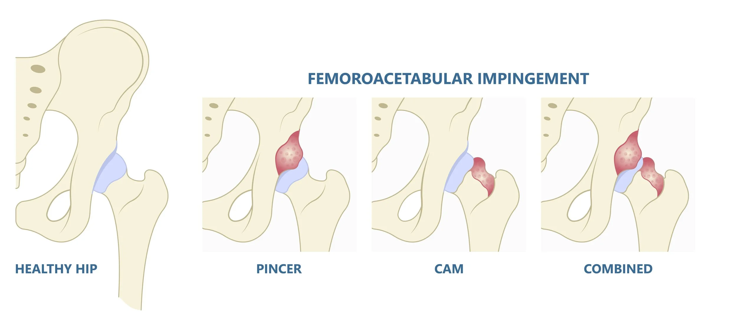
The hip is a ball-and-socket joint. In a normally shaped hip, the ball rotates smoothly within the socket through a full range of movement. In FAI, the geometry of one or both surfaces is abnormal - creating a zone where the bones clash as the hip moves through certain ranges.
FAI is not a disease and is not typically the result of a single injury. It reflects the shape of the bone - in many cases shaped by growth patterns during adolescence. Some people have this morphology and never develop symptoms. Others, particularly those who are physically active or who spend long periods in hip flexion, develop pain as the repetitive abnormal contact takes its toll on the joint over time.
Types of FAI
There are two main types of FAI, which frequently occur together.
Cam Impingement
Cam impingement involves extra bone at the junction of the femoral head and neck, creating a non-spherical shape to the ball. As the hip flexes, this extra bone engages the rim of the socket, shearing the labrum and damaging the cartilage just inside the acetabular rim. Cam morphology is more common in men and is frequently seen in individuals who were highly active in sport during adolescence, particularly activities involving repeated hip flexion such as football, rugby, and cycling.
Pincer Impingement
Pincer impingement involves over-coverage of the femoral head by the acetabulum; the socket covers too much of the ball. As the hip moves, the labrum is pinched between the rim of the socket and the femoral neck. Pincer morphology is more common in women and is often associated with a deeper or retroverted (backwards-tilted) acetabulum.
Mixed (Combined) FAI
Most patients with symptomatic FAI have elements of both cam and pincer morphology. This is referred to as mixed or combined FAI, and it is the most common pattern seen in clinical practice.
What Causes FAI?
FAI is largely determined by bone shape, which develops during growth. The exact cause is not fully understood, but several factors appear to increase the likelihood of developing symptomatic FAI:
High levels of sport participation during adolescence - particularly activities involving repeated hip flexion and rotation (football, rugby, gymnastics, cycling, dance)
Male sex - cam morphology is significantly more common in men
Hip conditions in childhood - conditions such as Perthes disease or slipped capital femoral epiphysis (SCFE) can alter the shape of the femoral head and predispose to FAI in adult life
Family history - there is evidence of a genetic component to hip morphology
It is important to note that having cam or pincer morphology on imaging does not automatically mean symptoms will develop. Many people with FAI morphology live without pain. Symptoms develop when the impingement is significant enough, and the demands placed on the hip exceed what the joint can comfortably accommodate.
Symptoms
The characteristic symptom of FAI is deep groin pain - located at the front of the hip, deep within the joint. Patients often describe what clinicians call the C-sign: they cup their hand around the front of the hip, with the thumb towards the back and the fingers pointing forward, when indicating where the pain is. This location is distinct from the outer hip pain associated with trochanteric bursitis, or the buttock pain more typical of lumbar spine referral.
Pain is typically:
Worse with prolonged sitting, particularly with the hip flexed beyond 90 degrees
Provoked by getting up from a low chair or out of a car
Present with pivoting, squatting, or hip rotation activities
Aggravated by impact sport or sustained physical activity
Associated with a click, catch, or giving-way sensation in some patients
FAI is frequently misdiagnosed in primary care as a hip flexor strain, a groin strain, or referred pain from the lumbar spine. Many patients present having already completed one or more courses of physiotherapy directed at these structures without meaningful improvement. If groin or deep hip pain has persisted beyond three months despite good physiotherapy, further investigation is warranted.
Diagnosis
Clinical Assessment
Diagnosis begins with a clinical history and physical examination. The FADIR test (flexion, adduction, and internal rotation of the hip) reproduces pain in most patients with symptomatic FAI by recreating the zone of impingement. Reduced internal rotation relative to the other side is a common finding. The FABER test (flexion, abduction, and external rotation) may also be positive, particularly in pincer-type impingement.
Imaging
Plain x-ray is the appropriate first investigation and can be arranged by your GP. The standard views are an AP pelvis x-ray and a Dunn lateral view (with the hip in 45 degrees of flexion). On these views:
Cam morphology appears as a loss of the normal spherical offset between the femoral head and neck - quantified by the alpha angle, which is considered abnormal above approximately 55 degrees
Pincer morphology appears as a crossover sign (the anterior acetabular rim crosses the posterior rim on the AP view) or a coxa profunda (deepened socket)
X-ray does not show the labrum or articular cartilage directly. If the clinical picture and x-ray findings are consistent with FAI, an MRI arthrogram is the next step. This is an MRI scan performed after injection of contrast medium into the joint, which provides detailed images of the labrum and cartilage surfaces. In New Zealand, an MRI arthrogram is arranged through the specialist after referral - it is not typically requested in primary care.
Treatment
Not every patient with FAI requires surgery. Treatment decisions are guided by the severity of symptoms, the degree of structural abnormality on imaging, the patient's age and activity goals, and whether significant labral or cartilage damage is present.
Physiotherapy
Physiotherapy is the appropriate starting point for most patients with FAI. A targeted programme addressing hip abductor and external rotator strength, core and lumbopelvic stability, and movement pattern modification can reduce the forces transmitted through the impingement zone and meaningfully improve symptoms. For patients with mild structural changes and no significant labral or cartilage damage, physiotherapy alone can be an effective long-term management strategy.
Activity Modification
Identifying and reducing activities that consistently provoke impingement, particularly sustained deep hip flexion, heavy squatting, or high-impact pivoting sport, can help manage symptoms while physiotherapy is progressing. This does not mean permanent activity restriction, but a period of load management while the hip is being rehabilitated.
Hip Arthroscopy for FAI
For patients whose symptoms have not improved adequately after a reasonable trial of conservative management (typically three to six months) and in whom structural pathology is confirmed on imaging, hip arthroscopy is the appropriate surgical option.
Hip arthroscopy for FAI is a keyhole procedure performed under general anaesthetic, usually as a day surgery. Through two or three small incisions around the hip, a camera and fine instruments are introduced into the joint. The procedure typically addresses:
Cam resection (femoral osteoplasty) - the extra bone at the femoral head-neck junction is precisely reshaped to restore the normal spherical contour of the femoral head
Pincer correction (acetabular osteoplasty) - where the socket is over-covering, the rim is trimmed to reduce the impingement
Labral repair - the damaged labrum is repaired and re-anchored to the rim of the socket using suture anchors. In most cases the labrum can be preserved and repaired rather than removed
The procedure typically takes 60 to 120 minutes. Addressing the underlying bony morphology at the time of labral repair, not just the labral damage itself, is important for reducing the risk of recurrence.
Recovery
Recovery from hip arthroscopy for FAI follows a structured rehabilitation programme, and the timeline varies depending on the findings at surgery and the demands of the patient's activities.
In the first few weeks, crutches are used to protect the hip while the labral repair heals. Physiotherapy begins early - gentle range of motion exercises in the first two weeks, progressing to strengthening as healing allows. Most patients are walking without crutches within two to four weeks.
Return to desk work and light activity is generally around four to six weeks. Return to higher-impact activity and sport varies depending on the demands involved, but is typically in the range of three to six months. Strength and movement quality are assessed throughout rehabilitation to guide return-to-activity decisions.
The aim of surgery is to correct the structural cause of the impingement, repair the labrum, and prevent further joint damage. Symptoms typically improve progressively over the months following surgery, and rehabilitation continues throughout this period.
Getting a Referral
If you have symptoms consistent with FAI, the first step is to see your GP or physiotherapist. They can arrange the initial x-rays (AP pelvis and Dunn lateral view) and provide a referral for specialist assessment if the clinical picture warrants it.
This guide is intended for general patient education and does not replace an individual consultation. If you have concerns about hip pain, please speak with your GP, physiotherapist or other health professional.