
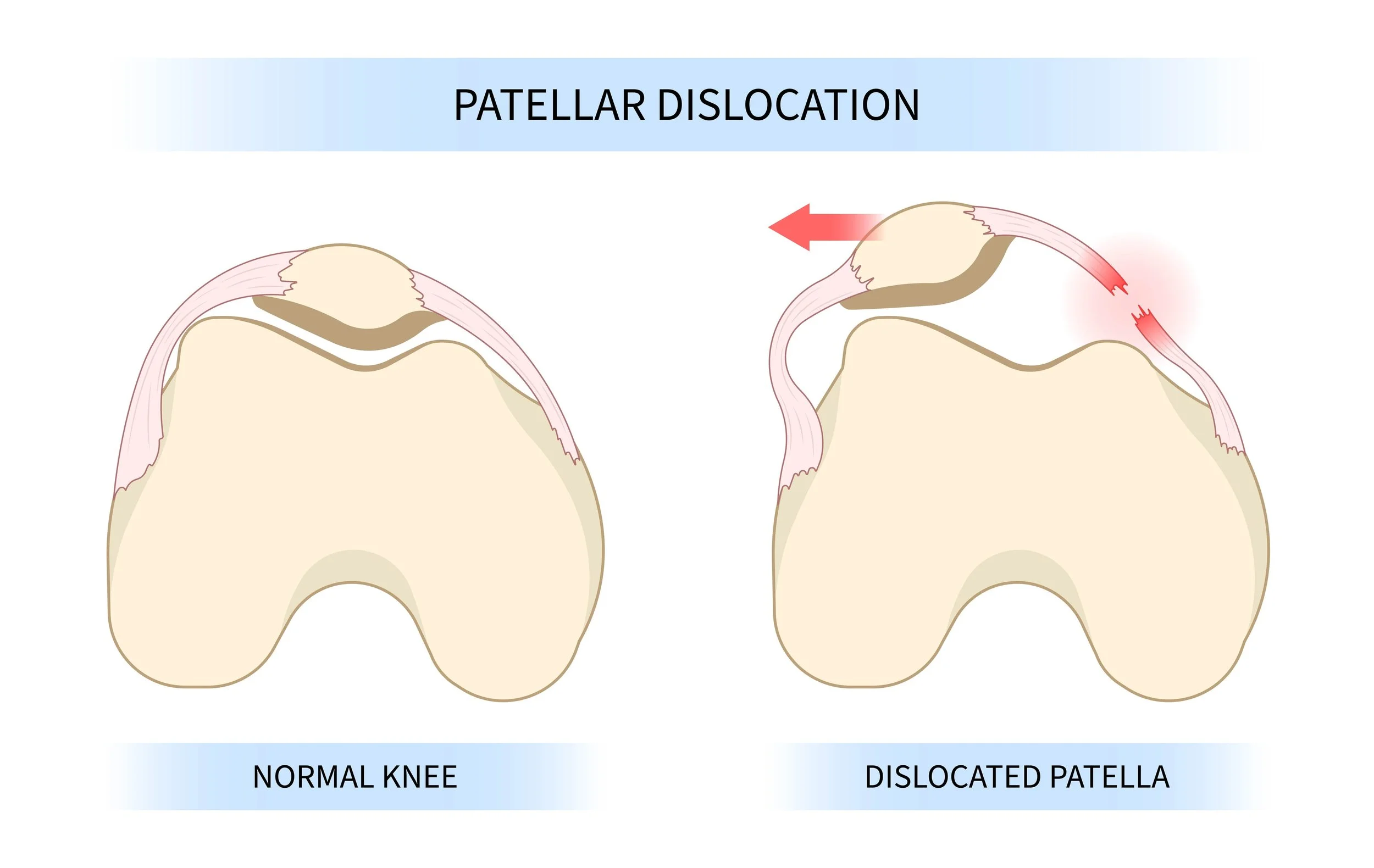
The patella (kneecap) is designed to sit in a groove at the front of the knee called the trochlea, gliding smoothly up and down as the knee bends and straightens. In some people, the patella is prone to sliding out of that groove. This is called patellar instability, and it ranges from a feeling of unsteadiness in the knee to full dislocation, where the patella slips completely to the outside of the joint.
For many patients, instability is not just an acute injury - it is an ongoing problem driven by anatomy. Understanding what causes the patella to dislocate, and why it tends to happen again, is central to choosing the right management.
What Causes Patellar Instability?
Patellar instability is usually the result of one or more anatomical factors that reduce the natural stability of the patellofemoral joint:
Trochlear dysplasia
The trochlear groove is the channel in the femur that guides the patella. In some people, this groove is shallow or poorly formed; a condition called trochlear dysplasia. A shallow groove provides less mechanical containment, making it easier for the patella to slip laterally (outwards).
Patella alta
Patella alta means the patella sits higher than normal relative to the trochlear groove. Because the patella engages with the groove later during knee flexion, it is less well-contained at the beginning of movement - the point when dislocation is most likely to occur.
MPFL insufficiency
The medial patellofemoral ligament (MPFL) is the primary soft-tissue restraint that prevents the patella from sliding outward. It runs from the medial (inside) edge of the patella to a point on the inner side of the femur. In virtually every acute patellar dislocation, the MPFL is torn or stretched. If it heals in a lengthened position, or does not heal adequately, the patella remains prone to further instability.
Increased TT-TG distance
The tibial tubercle (the bony prominence at the top of the shin) is where the patellar tendon attaches. If this is positioned too far to the outside relative to the centre of the trochlear groove, the patella is pulled laterally during knee extension. This is measured on MRI or CT imaging and is an important factor in surgical planning.
Symptoms
Patellar instability presents differently depending on whether a dislocation has occurred and how frequently it happens:
After a first dislocation, the knee is typically swollen and painful. The kneecap usually relocates spontaneously or is relocated before the patient seeks medical attention. There is often tenderness on the inner aspect of the knee where the MPFL has torn.
Between episodes, common symptoms include:
A feeling that the knee might "give way," particularly when pivoting, twisting, or changing direction
Pain around and behind the patella, especially on stairs or after prolonged sitting
Swelling after activity
Apprehension - a sensation of unsteadiness when the knee is put in positions associated with previous dislocation
In patients with recurrent instability, the threshold for dislocation often decreases over time, and episodes may occur with progressively less force. Each dislocation carries a risk of damage to the cartilage on the posterior surface of the patella and the front of the femur.
Diagnosis
Diagnosis involves a combination of clinical examination and imaging.
Clinical examination
Assessment typically includes evaluation of patellar position and tracking through range of motion, and specific tests including the patellar apprehension test - where gentle lateral pressure on the patella reproduces the patient's anxiety about dislocation. Lower limb alignment and muscle strength are also assessed.
Imaging
Plain x-rays of the knee - including an AP, lateral, and skyline (axial) view - provide the first level of assessment. The lateral view allows measurement of patella height, and the skyline view gives information about the shape of the trochlear groove.
MRI confirms the status of the MPFL, identifies any cartilage damage inside the joint, and provides additional detail on trochlear morphology. CT scanning or MRI is used when precise measurement of the TT-TG distance is required for surgical planning.
Treatment
Physiotherapy
For a first episode of patellar dislocation in a patient without significant anatomical risk factors, physiotherapy is appropriate initial management. A structured programme focuses on strengthening the muscles around the knee - particularly the VMO (vastus medialis obliquus), the inner portion of the quadriceps - as well as the hip abductors and external rotators, which influence the position of the femur and patella during movement. Taping or bracing may provide additional support during rehabilitation.
Physiotherapy alone may not be sufficient in patients with recurrent instability, significant anatomical abnormalities, or cartilage damage at the time of dislocation.
MPFL Reconstruction
When instability recurs despite physiotherapy, or when anatomical features make recurrence very likely, surgical reconstruction of the MPFL is typically recommended.
MPFL reconstruction involves replacing the torn or insufficient ligament with a graft, most commonly taken from the gracilis tendon behind the knee or the quadriceps tendon at the front. The graft is fixed to the medial edge of the patella and to the anatomical footprint of the MPFL on the medial femur, restoring the restraint that prevents the patella from dislocating laterally.
The procedure is performed under general anaesthetic and is typically undertaken as day surgery.
Additional procedures
In some patients, bony anatomy is a significant contributing factor and needs to be addressed alongside MPFL reconstruction.
Tibial tubercle transfer (TTT) is indicated when the TT-TG distance is elevated or when patella alta is present - and in many cases both factors coexist. The tibial tubercle is the bony prominence at the front of the shin where the patellar tendon attaches. When the TT-TG distance is elevated (typically above 20 mm), the tubercle is moved medially to reduce the lateral pull on the patella. In patella alta, where the patella sits too high relative to the trochlear groove, distalisation of the tubercle - moving it downward - brings the patella into earlier engagement with the groove, improving containment at the beginning of knee flexion where the risk of dislocation is greatest. Correcting patella alta through distalisation is often as important as, or more important than, medialisation when both deformities are present, and the direction of transfer is tailored to the individual's anatomy. The repositioned fragment is fixed with screws and heals in its new position over several weeks, which extends the overall recovery timeline compared with an isolated MPFL reconstruction.
Other anatomical factors can contribute to patellar instability even when they are not directly corrected surgically. Increased femoral anteversion - excessive inward rotation of the femur - effectively displaces the trochlear groove medially relative to the tibial tubercle, increasing the functional lateral force on the patella. Genu valgum (knock-knee alignment) similarly predisposes the patella to lateral tracking. These factors are identified and assessed as part of a thorough preoperative workup, and they inform the overall treatment plan, the choice of procedures, and the expectations for rehabilitation.
The appropriate combination of procedures is determined by the individual's anatomy and planned on a case-by-case basis. In straightforward cases, MPFL reconstruction alone is sufficient. Where significant bony contributions are present, additional procedures may be performed in the same operative session.
Recovery
Recovery after MPFL reconstruction follows a phased rehabilitation programme. The timeline below reflects an isolated MPFL reconstruction; where additional procedures such as tibial tubercle transfer are performed, recovery is typically longer as bone healing must occur before full rehabilitation can progress.
In the first two weeks, crutches are used for comfort and the knee is protected. Ice, elevation, and gentle range-of-motion exercises are begun early to limit swelling and maintain mobility. Most patients are walking comfortably without crutches by six weeks and have recovered a useful range of motion.
By three months, low-impact activities such as cycling and swimming are generally appropriate. Strengthening continues with a progressive focus on quadriceps and hip muscle control. By four to six months, jogging and light sport-specific training may be introduced depending on progress and individual goals.
Return to pivoting or contact sport is typically in the range of six to nine months for those undertaking a full rehabilitation programme. Clearance from both the treating surgeon and physiotherapist is required before return to sport. Strength and movement quality are assessed throughout rehabilitation to guide this decision.
Recovery timelines vary between individuals and depend on the procedures performed, the extent of any cartilage damage present at surgery, and adherence to rehabilitation. The timeframes above represent typical ranges rather than guaranteed outcomes.
Getting a Referral
If you have experienced one or more episodes of patellar dislocation, or have ongoing symptoms of instability, your GP or physiotherapist can refer you to an orthopaedic surgeon for assessment - they will typically arrange plain x-rays of the knee before referral.
If your instability is related to an injury - a fall, a collision during sport, or another traumatic event - the assessment and any subsequent treatment may be eligible for ACC cover. Your GP or physiotherapist can assist with the ACC referral process.
Key Points
A first dislocation always tears the MPFL
The medial patellofemoral ligament is injured in virtually every acute patellar dislocation. The question is whether it heals adequately - if it does not, the patella remains vulnerable to further instability.
Physiotherapy is first-line for a first episode
In patients without significant anatomical risk factors, a structured strengthening programme targeting the VMO and hip stabilisers is the appropriate starting point and can be effective long-term management.
MPFL reconstruction is the core surgical procedure
When surgery is indicated, reconstruction of the MPFL restores the primary soft-tissue restraint to lateral patellar translation. A graft from the gracilis (hamstring) tendon is used to replace the damaged ligament.
Bony procedures are added when anatomy requires it
If the TT-TG distance is elevated, or the patella sits too high (patella alta), a tibial tubercle transfer corrects the line of pull of the patellar tendon and resting position of the patella. If there is severe femoral malalignment, a derotational osteotomy may be required. The appropriate combination is planned individually.
Recovery to pivoting sport takes nine to twelve months
A full rehabilitation programme is essential. Return to sport is guided by strength and movement quality assessments rather than time alone, and requires clearance from both surgeon and physiotherapist.
This article is for general educational purposes only and does not constitute medical advice. Please consult your GP, physiotherapist, or a specialist for advice about your individual circumstances.